
|Articles|May 1, 2005
Advances in Imaging for Diagnosis of Alzheimer Disease
Author(s)C. P. Kaiser
Often, a clinical diagnosis of AD comes too late for an individual to benefit from treatment. Clinicians can assign the diagnosis of mild cognitive impairment (MCI) to patients with memory complaints, but they cannot state emphatically which of these individuals will progress to AD. Typically, about 10% to 15% of persons diagnosed with MCI convert to AD within a year, while 30% to 40% do not convert--at least not for another 6 to 8 years.
Advertisement
A woman with Alzheimer disease (AD) dies, and her adult children discuss undergoing a test to determine whether they carry the apolipoprotein E4 (ApoE4) allele, a common AD susceptibility gene. As worrisome as a positive test result would be, these family members will have more certainty about AD risk than the thousands of noncarriers who present to family practitioners with memory loss and impaired thinking.
Often, a clinical diagnosis of AD comes too late for an individual to benefit from treatment. Clinicians can assign the diagnosis of mild cognitive impairment (MCI) to patients with memory complaints, but they cannot state emphatically which of these individuals will progress to AD. Typically, about 10% to 15% of persons diagnosed with MCI convert to AD within a year, while 30% to 40% do not convert--at least not for another 6 to 8 years.1
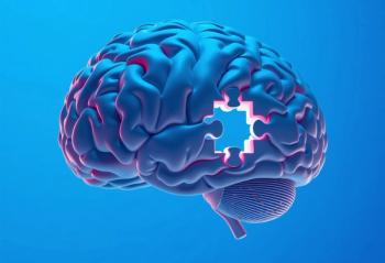
"Patients, families, and even physicians are in a state of suspended animation. We don't know whether we should start treating for AD or tell the family to look into assisted living," said Norman L. Foster, MD, professor of neurology at the University of Michigan Medical School in Ann Arbor. As people live longer, clinicians need clear markers that can identify earlier and more accurately those at risk for developing sporadic AD, which is different from and more prevalent than the familial type of AD. Diagnostic imaging plays an increasingly crucial role in detecting AD, distinguishing it from other debilitating dementias, and monitoring the progress of drug therapy.
A range of imaging techniques has been developed that enable researchers to visualize different features of AD, notably volumetric changes measured with MRI and metabolic changes measured with positron emission tomography (PET) and single photon emission computerized tomography (SPECT). Although these techniques are sensitive to AD pathology, they lack specificity. Efforts are under way to combine these tests with use of biomarkers, such as protein and enzyme expression, and other imaging techniques, such as functional MRI and MR spectroscopy (MRS), to increase specificity.
In October, the National Institute on Aging announced the Alzheimer Disease Neuroimaging Initiative, a 5-year $60 million project that seeks to recruit 800 participants to determine the best combination of imaging, biologic marker assessments, and clinical and neuropsychological assessments to detect MCI and AD. For information about the initiative, go to www.alzheimers.org/nianews/
nianews70.html.
Imaging will include serial MRI, and the study will be the first setting for serial fluorodeoxyglucose (FDG) PET studies. Recruitment for the clinical trial had not begun at press time but was scheduled to begin this spring at 50 sites (www.clinicaltrials.gov/ct/show/NCT00106899?order=1). In general, imaging is needed to address 3 unanswered clinical questions:
*How can mild forgetfulness caused by early, predementia AD be distinguished from mild forgetfulness caused by normal aging? The answer to this question will be important in finding strategies for early detection.
*How can dementia caused by AD be distinguished from other dementing illnesses? This is a question related to diagnostic specificity.
*What is the best way to test for drug efficacy? This question is important both for drug development and for following the course of approved drugs.
FOCUSING ON the amyloid CASCADE
Many researchers consider histologic imaging to be the future of dementia detection. This involves the use of novel radiotracers that bind to specific brain lesions associated with AD pathology, such as amyloid plaques and neurofibrillary tangles.
The "amyloid cascade" is a widely accepted theory postulating that the primary cause of AD is a series of events in the brain brought about by amyloid in various forms. A minority view holds that amyloid is simply a by-product of other processes, such as oxidative damage, inflammation, or cholesterol buildup. In any case, amyloid seems to play a role in AD pathology. Diagnostic imaging techniques--and treatment strategies--are focusing on amyloid.
Most drug companies are bet- ting on the cascade theory, according to Chet Mathis, PhD, a professor of radiology and pharmaceutical sciences at the University of Pittsburgh. The university has licensed the technology for 2-(4-methylaminophenyl)-6-hydroxybenzothiazole, an agent known as Pittsburgh compound B,2 or "PIB," to Amersham, which was recently purchased by GE Healthcare. GE intends to work with drug companies to use PIB, which binds to b-amyloid plaques in the brain, to monitor antiamyloid therapies in clinical trials. Mathis and PIB codeveloper William E. Klunk, MD, PhD, associate professor of psychiatry at the University of Pittsburgh, are developing a fluorine-18-labeled analog of PIB that would give the tracer a 2-hour half-life, making it more clinically practical than its current carbon-11 cousin, which
has a 20-minute half-life.
Research has shown that PIB results in a much greater retention rate in patients with AD and MCI than in healthy controls. But an overlap between those with disease and controls leads to more questions, Klunk said last July at the International Conference on Alzheimer Disease and Related Disorders (ICAD). "Are these false-positives or are they cognitively normal people with deposits of amyloid plaques? Does an initial deposit of amyloid eventually lead to AD? These are questions we are trying to answer," he said.
Investigators at the University of California, Los Angeles, developed 18-fluorine-dimethylami- no-dicyano-naphthalene propene (FDDNP), a PET tracer that binds to neurofibrillary tangles and b-amyloid plaques.3 Initial results with FDDNP indicate a strong correlation between the PET signal and the degree of memory loss, according to Daniel H. Silverman, MD, PhD, head of neuronuclear imaging at the institution, who also spoke at ICAD.
The distribution volumes of FDDNP showed a 15% to 40% increase in neurofibrillary tangles and b-amyloid plaques, depending on disease progression, in patients with AD compared with age-matched controls. Areas of increased FDDNP uptake correlate inversely with FDG hypometabolic patterns. Furthermore, FDDNP binding was associated with decreased neuron density in the hippocampus of patients with AD. Silverman echoed Mathis when he emphasized that the most promising use of histologic imaging may be to evaluate and monitor the efficacy of antiamyloid drugs.
Researchers at the Hospital of the University of Pennsylvania in Philadelphia have developed 4- N-methylamino-49-hydroxystibene (SB-13), a C-11-labeled tracer that binds to b-amyloid plaques.4 SB-13 showed results similar to those of PIB in a small study comparing the 2 tracers in 6 patients with AD and 6 healthy controls. These data have been presented at international conferences, said Daniel Skovronsky, MD, PhD, a neuropathologist at the University of Pennsylvania. His team worked with researchers from the University of Toronto. "It strengthens the case that both compounds are recognizing amyloid plaques," Skovronsky said.
Futhermore, a team led by Hank Kung, PhD, professor of radiology and pharmacology at the University of Pennsylvania, has developed 6-iodo-2-(49-dimethylamino-)phenyl-imidazo[1,2] pyridine (IMPY), an iodine-123-labeled compound for SPECT imaging of b-amyloid plaques.4 They have accumulated promising animal data and expect to have human data soon, which should indicate whether the low resolution of SPECT is sufficient to reveal the burden of amyloid plaque, Skovronsky said. The researchers want to give the nearly 5 million patients with AD the option to have SPECT imaging, since it is more widely available than PET.
EXAMINING GENETIC RISK
Researchers at the University of Arizona have been detecting and tracking brain changes that precede the onset of memory and thinking problems in persons with the ApoE4 allele. They hope to use imaging to test promising primary prevention therapies--treatment before symptoms occur--without needing to study thousands of research volunteers or wait many years to determine whether or when research subjects develop symptoms, according to Eric M. Reiman, MD, clinical director of neurogenomics at the Translational Genomics Research Institute in Phoenix and a professor of psychiatry at the University of Arizona.
Reiman cites the Women's Health Initiative Memory Study,5 an ancillary study of the Women's Health Initiative, as an example of the "old-fashioned" type of study that takes many years and millions of dollars to complete. The Women's Health Initiative was halted in June 2002 because evidence emerged that combined estrogen and progestin hormonal therapy increased the risk of cardiovascular disease and breast cancer. Preliminary results of the memory component, which used cognitive tests to gauge memory and thinking, revealed that hormone replacement therapy increased the risk of developing dementia. The memory study included nearly 5000 women between the ages of 65 and 80 who agreed to participate in a 10-year placebo-controlled trial.
"What would have happened if investigators had used hormone replacement therapy as a primary prevention therapy soon after menopause? Such a study would require 50,000 subjects and take more than 20 years," Reiman commented. "Most drug companies' patents would end before the study's conclusion."
This is where imaging comes in. The Arizona researchers have been longitudinally studying 200 people, aged an average of 55 years, who have either 2 copies, 1 copy, or no copy of the ApoE4 gene. Every 2 years, participants undergo MRI scanning, PET imaging, and a battery of memory tests. The researchers already have detected some of the same brain changes in asymptomatic carriers of the gene as those seen in patients with AD. They have noted a 2-year decline in these measurements, similar to that seen in patients with dementia. Based on these data, the investigators estimate that they would need 100 individuals over the course of 2 years to test a promising primary prevention therapy using imaging analysis rather than cognitive tests as the primary end point.
GlaxoSmithKline is one of the first drug companies to take advantage of Reiman's theories. The company has sponsored a 1-year "proof of concept" study by using FDG PET to determine whether the drug rosiglitazone maleate (Avandia), which is approved for type 2 diabetes, can slow the progression of AD. Imaging analysis, rather than cognitive testing, is the primary end point. "With many more treatments coming on line, it's not going to be possible to spend years and millions of dollars testing each one," Reiman said.
MRI: OLD AND NEW
In unaffected persons genetically at risk for early-onset AD, whole-brain atrophy measured with MRI has been shown to predict decline to AD. In a recent study, Clifford R. Jack, Jr, MD, a professor of diagnostic radiology at the Mayo Clinic in Rochester, Minn, and colleagues6 compared hippocampal volume and the rate of change in the whole brain in normal elderly controls and patients with MCI. The controls who converted to MCI had smaller volumes at baseline.
The percentage of people who convert from normal to AD is typically small, about 1% a year. Thus, even though Jack's research shows specific and overall volume changes associated with MCI and AD, it is not specific for the disease. It becomes an engineering question, he said. "We know the brain shrinks in patients with AD and also in people who are in the process of developing AD. But can we make precise measurements of that atrophy? If we can, then even very small changes can give us a diagnosis," Jack said.
One technique that promises to improve diagnostic specificity is arterial spin-labeled perfusion MRI.7 The procedure, which has been used in stroke treatment, can be performed in 10 minutes with a commercial scanner. The MR sequence inverts the polarity of water protons, which then flow into the region of interest and perfuse into extravascular space. The inverted protons, mixing with protons of a different polarity, are readily distinguished. Studies using this sequence in persons with MCI and AD have demonstrated hypoperfusion similar to that seen in FDG-PET and hexamethylpropyleneamine oxine SPECT.8
Patients with suspected dementia will typically undergo structural MRI scanning. They may then be referred for other imaging techniques such as SPECT or PET. An advantage of the arterial spin-labeled technique is that a structural MRI scan and a perfusion scan can be obtained at the same time, said Norbert Schuff, PhD, an associate professor of radiology at the San Francisco Veterans Affairs Medical Center.
MRS also holds promise as a tool that can add specificity to a diagnosis of AD or MCI. Researchers have shown that patients with AD exhibit unique metabolite spectra. The most promising marker for differentiating normal healthy subjects from patients with MCI and AD is the ratio of myo-inositol (MI) to N-acetylaspartate (NAA). Many groups have consistently found that NAA, a marker unique to neuronal tissue, is reduced and MI is increased in patients with AD. Several groups have also found reduced NAA and increased MI in those with MCI. The Mayo Clinic team found that MI is increased earlier than NAA is reduced. This could mean that MI is a more sensitive marker than NAA for early detection of MCI.
Performing MRS can be challenging, Schuff said, but newer automated programs will reduce the degree of operator expertise currently needed. Higher-field scanners also will be more sensitive to some metabolites, increasing the value of MRS.
Blood oxygen level-dependent (BOLD) functional MRI may have a place in diagnosing MCI or AD and in determining the efficacy of treatment, but BOLD imaging can be noisy. Noisiness and other variables differ between patients and between scans from the same patient. Serge Rombouts, PhD, a researcher at the VU University Medical Center in Amsterdam, is trying to overcome these variables by using BOLD imaging during the resting stage.9 "It's very reproducible, like an EEG," he said.
Rombouts and colleagues have scanned a small number of healthy controls and have found that the networks that show BOLD activity during the resting stage are the same networks, such as those of visual cortex and the motor cortex, that show activation during different memory tasks. BOLD MRI also has recorded the effects of medication in subjects with MCI and AD.
RECOGNIZED VALUE OF AN OLD TECHNIQUE
A paper by Philip Scheltens, MD, PhD, and coworkers,10 published in 1992 in the Journal of Neurology, Neurosurgery & Psychiatry, is receiving a great deal of attention today. The study details a way to roughly estimate hippocampal atrophy based on MR coronal slices. Scheltens, a neurologist at the VU University Medical Center, has found, in studying several different population groups, that visual assessment of atrophy has a high accuracy for predicting who will convert. "Despite the advancement of volumetric and whole-brain analysis techniques, which can take hours, this old-fashioned way is still relevant for diagnostic purposes and for predicting who will convert from MCI to AD," Scheltens said.
Visual assessment takes just a few seconds for an experienced rater. It is convenient for large clinical trials that can include hundreds of scans. Raters score the left and right side of the brain separately. Scores of 0 and 1 are associated with normalcy; scores of 2, 3, and 4 are associated with disease. In a comparative study, visual analysis predicted disease better than volumetric analysis. Unlike volumetric analysis, visual assessment takes a global view of the hippocampal region, including medial temporal atrophy, which is probably more sensitive to AD in patients with MCI, he said.
Of course, volumetric analysis is fast becoming operator-independent. The future will be in those automatic methods, Scheltens said. *
references
1. Solfrizzi V, Panza F, Colacicco AM, et al. Vascular risk factors, incidence of MCI, and rates of progression to dementia. Neurology. 2004;63:1882-1891.
2. Klunk WE, Engler H, Nordberg A, et al. Imaging brain amyloid in Alzheimer's disease with Pittsburgh Compound-B. Ann Neurol. 2004;55: 303-305.
3. Silverman DH. Brain 18F-FDG PET in the diagnosis of neurodegererative dementias: comparison with perfusion SPECT and with clinical evaluations lacking nuclear imaging. J Nucl Med. 2004;45:594-607.
4. Kung MP, Hou C, Zhuang ZP, et al. Binding of two potential imaging agents targeting amyloid plaques in postmortem brain tissues of patients with Alzheimer's disease [published correction appears in Brain Res. 2005;1031:302]. Brain Res. 2004;1025:98-105.
5. Shumaker SA, Legault C, Rapp SR, et al. Estrogen plus progestin and the incidence of dementia and mild cognitive impairment in postmenopausal women: the Women's Health Initiative Memory Study: a randomized controlled trial. JAMA. 2003;289:2651-2662.
6. Kantarci K, Petersen RC, Boeve BF, et al. DWI predicts future progression to Alzheimer disease in amnestic mild cognitive impairment. Neurology. 2005;64:902-904.
7. Jahng GH, Song E, Zhu XP, et al. Human brain: reliability and reproducibility of pulsed arterial spin-labeling perfusion MR imaging. Radiology. 2005;234:909-916.
8. Johnson NA, Jahng GH, Weiner MW, et al. Pattern of cerebral hypoperfusion in Alzheimer disease and mild cognitive impairment measured with arterial spin-labeling MR imaging: initial experience. Radiology. 2005;234:851-859.
9. Rombouts SA, Stam CJ, Kuijer JP, et al. Identifying confounds to increase specificity during a "no task condition." Evidence for hippocampal connectivity using fMRI. Neuroimage. 2003;20: 1236-1245.
10. Scheltens P, Leys D, Barkhof F, et al. Atrophy of medial temporal lobes on MRI in "probable" Alzheimer's disease and normal ageing: diagnostic value and neuropsychological correlates. J Neurol Neurosurg Psychiatry. 1992;55:967-972.
A version of this article appeared in the December 2004 issue of Diagnostic Imaging. It has been revised for Applied Neurology.
C.P. Kaiser is a news editor for the journal Diagnostic Imaging. He is based in Philadelphia.
Functional MRI of traverse sections of the brain with color overlay representing significant activation during the observation of faces. The blue line in the graph shows the activated region of the occipital cortex in normal elderly persons responding to a face. The yellow shows activation in patients with mild Alzheimer disease. (Image provided by S. Rombouts.)FDG-PET scan (top) shows normal brain (left) and the typical hypometabolic glucose pattern in the medial and lateral temporal lobe (right) characteristic of Alzheimer disease (AD). MRI scan (bottom) shows a normal brain of a 76-year-old person and hippocampal atrophy in a 75-year-old patient with AD. (Images provided by M. de Leon.)Pittsburgh Compound B (PIB)-PET scans overlaid onto structural magnetic resonance (MR) scans show increased retention of PIB, a carbon-11 (C-11) radiotracer that binds to b-amyloid plaques. (Images provided by C. Mathis.)
Advertisement
Related Content
Advertisement


Latest CME


Advertisement
Advertisement