CME
Article
Psychiatric Times
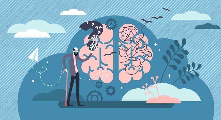
HIV-Associated Neurocognitive Disorder: An Updated Pathophysiology
Author(s):
In this CME, learn more about the pathophysiology of HIV-associated neurocognitive disorder and to review the research and various tools used to aid in the diagnosis.


gamjai/AdobeStock
CATEGORY 1 CME
Premiere Date: December 20, 2023
Expiration Date: June 20, 2025
This activity offers CE credits for:
1. Physicians (CME)
2. Other
All other clinicians either will receive a CME Attendance Certificate or may choose any of the types of CE credit being offered.
ACTIVITY GOAL
The goal of this activity is to explain the pathophysiology of HIV-associated neurocognitive disorder (HAND) and to review the research and various tools used to aid in the diagnosis of HAND.
LEARNING OBJECTIVES
After engaging with the content of this CME activity, you should be better prepared to:
1. Understand the pathophysiology of HAND.
2. Recognize the various tools used to aid in the diagnosis of HAND as well as the specific findings seen.
TARGET AUDIENCE
This accredited continuing education (CE) activity is intended for psychiatrists, psychologists, primary care physicians, physician assistants, nurse practitioners, and other health care professionals who seek to improve their care for patients with mental health disorders.
ACCREDITATION/CREDIT DESIGNATION/FINANCIAL SUPPORT
This activity has been planned and implemented in accordance with the accreditation requirements and policies of the Accreditation Council for Continuing Medical Education (ACCME) through the joint providership of Physicians’ Education Resource®, LLC, and Psychiatric Times®. Physicians’ Education Resource, LLC, is accredited by the ACCME to provide continuing medical education for physicians.
Physicians’ Education Resource, LLC, designates this enduring material for a maximum of 1.5 AMA PRA Category 1 Credits™. Physicians should claim only the credit commensurate with the extent of their participation in the activity.
This activity is funded entirely by Physicians’ Education Resource, LLC. No commercial support was received.
OFF-LABEL DISCLOSURE/DISCLAIMER
This accredited CE activity may or may not discuss investigational, unapproved, or off-label use of drugs. Participants are advised to consult prescribing information for any products discussed. The information provided in this accredited CE activity is for continuing medical education purposes only and is not meant to substitute for the independent clinical judgment of a physician relative to diagnostic or treatment options for a specific patient’s medical condition. The opinions expressed in the content are solely those of the individual faculty members and do not reflect those of Physicians’ Education Resource, LLC.
FACULTY, STAFF, AND PLANNERS’ DISCLOSURES AND CONFLICT OF INTEREST (COI) MITIGATION
None of the staff of Physicians’ Education Resource, LLC, or Psychiatric Times or the planners or the authors of this educational activity have relevant financial relationship(s) to disclose with ineligible companies whose primary business is producing, marketing, selling, reselling, or distributing health care products used by or on patients.
For content-related questions, email us at PTEditor@mmhgroup.com; for questions concerning the accreditation of this CME activity or how to claim credit, please contact info@gotoper.com and include “HIV-Associated Neurocognitive Disorder: An Updated Pathophysiology” in the subject line.
HOW TO CLAIM CREDIT
Once you have read the article, please use the following URL to evaluate and request credit: https://education.gotoper.com/activity/ptcme23dec. If you do not already have an account with PER®, you will be prompted to create one. You must have an account to evaluate and request credit for this activity.
Human immunodeficiency virus (HIV) is a retrovirus that infects and destroys CD4 T cells. In the United States, HIV infection affects an estimated 1,189,700 individuals 13 years and older.1 When uncontrolled, HIV infection progresses to acquired immunodeficiency syndrome (AIDS), which has a large spectrum of presentations.2 This large spectrum includes a myriad of neuropsychiatric, motor, and sensory impairments that occurs when HIV enters the central nervous system (CNS) via 1 of the many theorized pathways. Once HIV enters the CNS, it may produce a host of neurocognitive symptoms known as HIV-associated neurocognitive disorder (HAND).2
Prior to the development of highly active antiretroviral therapy (HAART), as many as 30% of HIV-positive patients displayed the most severe neurocognitive symptoms.3 Today, 40% to 60% of HIV-positive patients suffer from some form of neurocognitive impairment related to HAND.3,4 In fact, HIV type 1 (HIV-1) infection is the most common cause of major neurocognitive disorder (NCD) in young adults in the United States.4 Risk factors of developing HAND include older age, low CD4 counts, comorbid hepatitis C virus (HCV) infection, cardiovascular conditions (eg, hypertension, stroke), hypercholesterolemia, and preexisting psychiatric illnesses (eg, bipolar disorders, depressive disorders, anxiety disorders, substance-related disorders).5
The implementation of HAART regimens has dramatically reduced the incidence of HAND; however, studies show that the prevalence of HAND continues to increase, with a greater number of patients presenting with milder symptoms over longer periods of time. This is attributable to the increased life expectancy of HIV-positive patients as HAART regimens improve.3 Further, prevalence of neurocognitive impairment is reduced in patients who started HAART early after being infected, and it is higher in those who started their regimen late and with low CD4 counts.6 This article reviews the most up-to-date literature regarding the pathophysiology of HAND and provides a brief overview of the clinical features of, diagnostic imaging findings of, and treatments for this medical condition.
Clinical Features
The Frascati Panel, a working group assembled by the National Institute of Mental Health and the National Institute of Neurological Diseases and Stroke, reviewed the 1991 diagnostic guide for HAND created by the American Academy of Neurology’s AIDS Task Force to update its clinical utility. During their review, they discovered multiple areas that impacted clinical utility, including7:
1. the number of domains of neurocognitive impairment that were not clearly defined;
2. the degree of neurocognitive impairment that was not specified;
3. the overlap in diagnostic criteria among categories of severity; and
4. the exclusion of milder forms of illness.
To address these issues, the panel refined the diagnostic classification of HAND and provided 3 distinct conditions:
1. asymptomatic neurocognitive impairment (ANI): the neurocognitive impairment does not interfere with everyday functioning;
2. HIV-associated mild neurocognitive disorder (MND): the neurocognitive impairment produces at least mild interference in daily functioning, evidenced by either:
- self-reported reduced mental acuity and inefficiency in work, homemaking, and/or social functioning; or
- observation by knowledgeable others that the individual has undergone at least mild decline in mental acuity with resultant inefficiency in work, homemaking, and/or social functioning; and
3. HIV-associated dementia (HAD): the cognitive impairment produces marked interference with day-to-day functioning (eg, work, home life, social activities).
HAND can be described as a progressive neurocognitive illness in which patients may progress through conditions that advance from normal neurocognitive status to ANI to MND and, finally, to HAD.5 They cannot migrate in a retrograde manner through the conditions—thus, if a patient meets the diagnostic criteria for ANI, MND, or HAD, the diagnosis cannot be stepped back to a previous, less impairing diagnosis. If an individual no longer meets criteria for their condition at any time, an in remission specifier may be added.7
In ANI, there is no overt neurocognitive dysfunction noted in day-to-day function, but impairment may be seen on the results of standardized neuropsychological testing. In MND, there may be subjective reports of declining mental acuity and/or objective reports from individuals involved in the patient’s life.7 Some notable symptoms include difficulties in concentration, declining memory, and impaired executive function. In HAD, more severe signs of psychomotor slowing, irritability, agitation, and depressed mood may be present. If left untreated, end-stage major NCD signs (eg, bedridden state, mutism, and/or incontinence) can develop. In extremely severe cases, focal and generalized seizures can occur.6
Case 1
A 56-year-old woman with a psychiatric history of major depressive disorder (MDD), cannabis use disorder, cocaine use disorder, alcohol use disorder, and major NCD and a systemic medical history of HIV infection, asthma, HCV infection, hypertension, and Graves disease (which progressed to hypothyroidism after treatment) was brought to the emergency department after falling at home due to generalized weakness that worsened over the previous few weeks.
The patient had a history of poor compliance with HAART, as evidenced by multiple absolute CD4 counts below 200 cells/µL. Upon admission, her absolute CD4 count was less than 20 cells/µL, and viral loads for HIV and HCV were 307,000 copies/mL and 2.56 million copies/mL, respectively. She was admitted to the medical floor for further evaluation of weakness, but she quickly became uncooperative, agitated, and aggressive, cursing at staff and refusing all care. She requested to leave against medical advice, leading to a consultation with the consultation- liaison (CL) psychiatry team. On evaluation, the patient appeared disheveled, malodorous, agitated, and aggressive; she exhibited paranoia and said that the treatment team was “playing games” with her. It was determined that she lacked the capacity to make medical decisions. Her cognitive score was not obtained at the time.
A CT scan of the brain showed generalized volume loss, chronic left thalamus and cerebellar infarction with encephalomalacia. An MRI of the brain was attempted for further evaluation, but the patient refused to undergo testing. The medical team attempted to continue the patient’s daily HAART regimen of darunavir, 800 mg/cobicistat, 150 mg/emtricitabine, 200 mg/tenofovir alafenamide, 10 mg. However, the patient was nonadherent. Instead, she was prescribed atovaquone, azithromycin, and fluconazole for prophylaxis of opportunistic infections; she had poor adherence to this treatment, as well. Psychiatrically, the patient was managed with olanzapine, 5 mg twice daily, which improved her paranoia and had a mild effect on her irritability. Due to her inability to care for herself and sustained subjective neurocognitive impairment, she was ultimately discharged to a nursing home.
Case 2
A 62-year-old man with a history of schizophrenia, MND, hypertension, diabetes mellitus, and HIV infection was brought to the psychiatric emergency department from his nursing home by emergency medical services staff due to disorganized behavior, including inappropriate touching of staff, talking to himself, not sleeping, wandering the halls at night, and pulling the fire alarm. He had no history of substance use. Upon arrival, he was initially calm but internally preoccupied and oddly related; he offered sporadic responses to questions, and he was unable to provide any meaningful information. He required inpatient psychiatric admission for psychiatric stabilization.
On the psychiatry unit, the patient displayed disruptive and disorganized behavior, including pacing the halls, intruding on other patients, and entering other patients’ rooms. He had multiple symptoms of subjective cognitive impairment that included poor memory, word-finding difficulties, social withdrawal, and isolation. Due to the onset of incoherent speech, he was sent for an MRI; the results showed mild to moderate patchy and confluent T2 fluid-attenuated inversion recovery (FLAIR) hyperintensities within the periventricular and subcortical white matter and a focal T2 FLAIR hyperintensity in the left dorsal medulla. The MRI also revealed mild parenchymal volume loss in the supratentorial region and mild to moderate parenchymal volume loss in the infratentorial region.
The patient was maintained on daily HAART with bictegravir, 50 mg/emtricitabine, 200 mg/tenofovir alafenamide, 25 mg. He was relatively adherent to therapy, as evidenced by his absolute CD4 count of 526 cells/µL and undetectable viral load. He was treated on the unit with citalopram, 10 mg/d; memantine, 5 mg twice daily; quetiapine, 25 mg at night; valproic acid, 250 mg twice daily; and haloperidol decanoate, 100 mg monthly. He showed significant improvement in his disorganization and aggressive behavior; he eventually was discharged, and he returned to the nursing home.
Pathophysiology
HIV is a retrovirus with a complex life cycle. After HIV is introduced to the host, it uses its 2 major envelope molecules—gp120 and gp41—to enter CD4 cells. In these cells, the virus uses the host cell’s reproductive hardware to start its reproduction cycle. The virus can disassemble, replicate, reassemble, and exit host cells without alerting the host immune system.8 During the early course of HIV infection, the virus can easily infect CD4 T cells, macrophages, and monocytes.6 These cells can cross the blood-brain barrier (BBB) by transendothelial leukocyte migration, granting HIV easy access to the brain parenchyma.9 This is supported by evidence of inflammatory cerebrospinal fluid (CSF) changes seen in the asymptomatic phase of HIV infection.6
The most crucial cell population to the pathogenesis of HAND is the monocyte, particularly the CD14+/CD16+ subset.10 When compared with other cells, this subset has increased CCR5 coreceptors; in HIV-positive patients, its concentration density increases from 10% to 40% of peripheral blood monocytes.10 When monocytes enter the CNS and differentiate into macrophages, they remain fixed in the CNS for an extended period. If the differentiated macrophage happens to be infected with HIV, it can act as a long-standing reservoir of HIV in the CNS, paving the way for chronic inflammation and immune activation within the CNS.5,10
HIV-infected macrophages, microglia, and astrocytes secrete neurotoxic viral proteins that activate other CNS immune cells. Once activated, these cells secrete proinflammatory cytokines (including tumor necrosis factors (TNFs), interleukins, and other chemokines) to help recruit more monocytes and jump start neuroinflammation. Once fully activated, immune cells secrete neurotoxic host factors that, with the viral proteins, results in demyelination, pruning, and neuronal loss.5,10
Neurotoxic HIV viral proteins also alter glutamate metabolism in 3 major ways.4,5,10,11 First, they inhibit glutamate reuptake by other immune cells. Second, they increase the secretion of glutamate from nerve endings. Third, they have a role in the phosphorylation of glutamate receptors, which potentiates excitotoxicity.4,5,10,11 Glutamate plays a major role in regulating such multiple neuropsychiatric functions as learning, memory, and neuroplasticity, which are impaired domains observed in HAND.12


FIGURE. Pathophysiology of HAND13,a
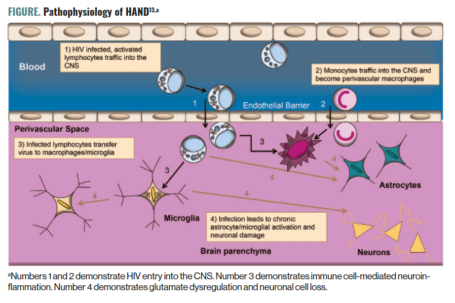
Based on the previously described mechanism, the stages of HAND pathophysiology can be conceptualized in 3 stages (Figure)13:
1. HIV entry into the CNS: HIV crosses the BBB via infected monocytes by transendothelial leukocyte migration5,6;
2. immune cell-mediated neuroinflammation: HIV-infected monocytes differentiate into macrophages and secrete neurotoxic viral proteins (these proteins act as a positive reinforcement mechanism to stimulate production of inflammatory cytokines, recruit more immune cells to the CNS, and potentiate neuroinflammation)5,10; and
3. glutamate dysregulation, excitotoxicity, and neuronal loss: viral and host neurotoxic proteins alter glutamate metabolism by inhibiting glutamate reuptake, increasing the secretion of glutamate, and phosphorylating glutamate receptors.5,10
The COVID-19 pandemic revealed new mechanisms to activate chronic diseases and displayed many of its own sequelae from COVID brain to COVID toes.14 COVID-19 also may exacerbate symptoms of HAND via synergistic pathways. Just as HIV can enter the CNS and cause demyelination and neuronal loss, COVID-19 can stimulate microglial activation and demyelination, leading to delirium. Another similarity noted in the literature includes COVID-induced hypercytokinemia leading to a neuroinflammatory response severe enough to cause disruption to the BBB and further recruitment of immune cells. COVID-induced neuroinflammation eventually leads to dysregulation of neurotransmitters (eg, glutamate dysregulation seen in HAND).15,16
Diagnostics
In the era of combination antiretroviral therapy, results from neuropsychiatric and neuropsychological testing do not always directly correspond with HIV load and CD4 count, highlighting the need for specific plasma and CSF biomarkers to predict prognosis and assist in the diagnosis of HAND. Modalities used to aid in the diagnosis of HAND include plasma studies, CSF analysis, and imaging.17
Plasma
Elevated levels of inflammatory cytokines and markers of immune activation (eg, IFN-α, IL-15, IP-10, TNFα, and MCP1) have been observed in patients with ANI and MND. Slow increases in IL-6, IL-8, IL-18, and IFNγ levels have been reported, whereas IL-10 levels tend to peak later.18 There is a strong correlation between MMP2 levels found in the plasma and CSF and changes in the structure and function of the brain.19 An increased plasma sCD14 level has been associated with impaired attention and concentration in patients with advanced disease and can serve as a potential biomarker to monitor progression of the disorder.20 Increased plasma levels of sCD163, a marker of activated monocytes, have been associated with neurocognitive impairment in HAND.21 Accumulations of bioactive lipids such as ceramide and sterol are seen in ANI and MND.22 Markers of neuronal injury like NFL protein, τ protein, and APP tend to appear later in the disease process; they are associated with low CD4+ T-cell counts.23,24
CSF
CSF biomarkers have not consistently demonstrated clinical usefulness in the diagnosis of HAND, but they may be helpful in identifying at-risk individuals. Neurocognitive impairment in HAND has been linked to elevated levels of cytokines (eg, IL-8, MCP1, granulocyte colony-stimulating factor, and IP-10) and markers of immune activation (eg, IL-6, CCL2).25,26 These biomarkers may represent a profile for HAND. In addition, markers of neuronal injury (eg, NFL, τ) may also be associated with more advanced cognitive impairment. Increased sCD14 level in the CSF has been linked to impaired neurocognition in HIV patients, suggesting that it may have potential as a biomarker for monitoring the progression of HAND.26
Imaging
As patients progress to the more advanced form of HAND, neuroimaging findings may become more prevalent. CT scans and MRIs are the initial neuroimaging modalities used to evaluate patients with HAND.27,28 Although no imaging findings are considered diagnostic for HAND, some common findings have been reported, which include27:
- gray-matter atrophy of the cerebral cortex;
- signal intensity and atrophy of deep white matter;
- loss of white matter integrity of the corpus collosum;
- bilateral periventricular and semiovale white matter lesions; and
- prominent cortical sulci and widened ventricle.
Other imaging modalities have been used to evaluate HAND; however, most are used in research and require more evidence for clinical use.27
Concluding Thoughts
The advent of HAART has done much to reduce the number of patients with HAND who present with severe neurocognitive impairment (particularly the HAD stage). Most affected patients now remain in the ANI to MND stages. It is prudent to continue reinforcement of HAART treatment and adherence to prevent the development of severe neurologic impairments and progression to HAD.
Significant progress has been made; however, further research could expand understanding of many domains. HIV can enter the CNS early in infection and pave the way for HAND development, and no prophylactic strategy for this is known. Additionally, many of the biomarkers and imaging modalities do not have sufficient evidence for clinical use.
Finally, HAART may minimize further cognitive decline by controlling HIV infection/AIDS, but we have no ability to treat the damage already done in patients with MND and HAD. Thus, it is imperative to expand our knowledge in all 3 domains of prevention, monitoring, and treatment.
It is no surprise that COVID-19, with all its interplay in other areas of medicine, has also demonstrated synergy with HIV infection in the progression of HAND. Further characterization of this relationship is needed to understand future steps in prevention, diagnosis, and treatment.
Consultation-liaison psychiatrists frequently are consulted to mitigate the sequelae of neurocognitive impairment, which may include insomnia, aggression, and psychosis, to name a few. This can lead health care professionals to have tunnel vision and address only the sequelae without considering the etiology. On the systemic medical side, it is not uncommon for internal medicine teams to conceptualize NCD as a psychiatric problem without considering HIV infection/AIDS or HAART regimen nonadherence as a potential factor in worsening NCD. Consultants must keep this possibility in mind when treating NCD patients, especially those with a history of HIV infection/AIDS.
Dr Garrels is PGY-4 and chief resident in the Department of Psychiatry at BronxCare Health System. Dr Kainth is PGY-1 at BronxCare Health System. Dr Zamiri is a psychiatry resident at BronxCare Health System. Dr Srinivas is a psychiatry resident at UAMS-Baptist Health and a geriatric telepsychiatry fellow at Michigan State University. Dr Laul is a psychiatry resident at the University of Miami. Dr Gunturu is director of the psychiatry residency training program at BronxCare Health System.
References
1. Statistics overview. CDC. Reviewed August 10, 2022. Accessed September 20, 2023. https://www.cdc.gov/hiv/statistics/overview/index.html
2. Alimonti JB, Ball TB, Fowke KR. Mechanisms of CD4+ T lymphocyte cell death in human immunodeficiency virus infection and AIDS. J Gen Virol. 2003;84(7):1649-1661.
3. Lindi KA, Marks DR, Kolson DL, Jordan-Sciutto KL. HIV-associated neurocognitive disorder: pathogenesis and therapeutic opportunities. J Neuroimmune Pharmacol. 2010;5(3):294-309.
4. Ghafouri M, Amini S, Khalili K, Sawaya BE. HIV-1 associated dementia: symptoms and causes. Retrovirology. 2006;3:28.
5. Saylor D, Dickens AM, Sacktor N, et al. HIV-associated neurocognitive disorder—pathogenesis and prospects for treatment. Nat Rev Neurol. 2016;12(4):234-248.
6. Eggers C, Arendt G, Hahn K, et al; German Association of Neuro-AIDS and Neuro-Infectionology (DGNANI). HIV-1-associated neurocognitive disorder: epidemiology, pathogenesis, diagnosis, and treatment. J Neurol. 2017;264:1715-1727.
7. Antinori A, Arendt G, Backer JT, et al. Updated research nosology for HIV-associated neurocognitive disorders. Neurology. 2007;69(18):1789-1799.
8. Simon V, Ho DD, Karim QA. HIV/AIDS epidemiology, pathogenesis, prevention, and treatment. Lancet. 2006;368(9534):489-504.
9. Takeshita Y, Ransohoff RM. Inflammatory cell trafficking across the blood-brain barrier (BBB): chemokine regulation and in vitro models. Immunol Rev. 2021;248(1):228-239.
10. Williams DW, Veenstra M, Gaskill PJ, et al. Monocytes mediate HIV neuropathogenesis: mechanisms that contribute to HIV associated neurocognitive disorders. Curr HIV Res. 2014;12(2):85-96.
11. Haughey NJ, Nath A, Mattson MP, et al. HIV-1 Tat through phosphorylation of NMDA receptors potentiates glutamate excitotoxicity. J Neurochem. 2001;78(3):457-467.
12. Huang Q, Xie Y, Hu Z, Tang X. Anti-N-methyl-D-aspartate receptor encephalitis: a review of pathogenic mechanisms, treatment, prognosis. Brain Res. 2020;1727:146549.
13. Gonzalez-Scarano F, Kolson DL. HIV associated neurocognitive disorder after the start of combination retroviral therapy. Psychiatric Times. 2019;36(9).
14. COVID toes, rashes: how the coronavirus can affect your skin. American Academy of Dermatology. Accessed March September 20, 2023. https://www.aad.org/public/diseases/coronavirus/covid-toes
15. Troyer EA, Kohn JN, Hong S. Are we facing a crashing wave of neuropsychiatric sequelae of COVID-19? neuropsychiatric symptoms and potential immunologic mechanisms. Brain Behav Immun. 2020;87:34-39.
16. Dantzer R. Neuroimmune interactions: from the brain to the immune system and vice versa. Physiol Rev. 2018;98(1):477-504.
17. Becker JT, Kingsley L, Mullen J, et al; Multicenter AIDS Cohort Study. Vascular risk factors, HIV serostatus, and cognitive dysfunction in gay and bisexual men. Neurology. 2009;73(16):1292-1299.
18. Ragin AB, Wu Y, Gao Y, et al. Brain alterations within the first 100 days of HIV infection. Ann Clin Transl Neurol. 2015;2(1):12-21.
19. Li S, Wu Y, Keating SM, et al. Matrix metalloproteinase levels in early HIV infection and relation to in vivo brain status. J Neurovirol. 2013;19(5):452-460.
20. Lyons JL, Uno H, Ancuta P, et al. Plasma sCD14 is a biomarker associated with impaired neurocognitive test performance in attention and learning domains in HIV infection. J Acquir Immune Defic Syndr. 2011;57(5):371-379.
21. Burdo TH, Weiffenbach A, Woods S, et al. Elevated sCD163 in plasma but not cerebrospinal fluid is a marker of neurocognitive impairment in HIV infection. AIDS. 2013;27(9):1387-1395.
22. Bandaru VVR, Mielke MM, Sacktor N, et al. A lipid storage-like disorder contributes to cognitive decline in HIV-infected subjects. Neurology. 2013;81(17):1492-1499.
23. Peterson J, Gisslen M, Zetterberg H, et al. Cerebrospinal fluid (CSF) neuronal biomarkers across the spectrum of HIV infection: hierarchy of injury and detection. PLoS One. 2014;9(12):e116081.
24. Krut JJ, Mellberg T, Price RW, et al. Biomarker evidence of axonal injury in neuroasymptomatic HIV-1 patients. PLoS One. 2014;9(2):e88591.
25. Yuan L, Qiao L, Wei F, et al. Cytokines in CSF correlate with HIV-associated neurocognitive disorders in the post-HAART era in China. J Neurovirol. 2013;19(2):144-149.
26. Kamat A, Lyons JL, Misra V, et al. Monocyte activation markers in cerebrospinal fluid associated with impaired neurocognitive testing in advanced HIV infection. J Acquir Immune Defic Syndr. 2012;60(3):234-243.
27. Haziot MEJ, Barbosa SP Jr, Vidal JE, et al. Neuroimaging of HIV-associated neurocognitive disorders. Dement Neuropsychol. 2015;9(4):380-384.
28. Thurnher MM, Schindler E, Thurnher SA, et al. Highly active antiretroviral therapy for patients with AIDS dementia complex: effect on MR imaging findings and clinical course. AJNR Am J Neuroradiol. 2000;21(4):670-678.


Newsletter
Receive trusted psychiatric news, expert analysis, and clinical insights — subscribe today to support your practice and your patients.