- Vol 34 No 3
- Volume 34
- Issue 3
Neuroanatomy and the 21st Century Psychiatrist
Why learn neuroanatomy? The goal for the physician is to be excitedly engaged in an ongoing process of expanding his or her knowledge about the brain and human behavior.
During most of medical history, all we knew about the brain was its gross anatomy. Then, in the late 1800s, the brain’s microscopic cellular structure began to be elucidated. Now, well into the 21st century, we also have remarkable insights into how the brain functions. Still, studying neuroanatomy is viewed as the first step in learning about the brain. Of course, this makes sense.
But, learning neuroanatomy is actually quite difficult, especially if you are a psychiatrist who is returning to this subject after having been away from the topic for some time. Therefore, the idea that one must first learn neuroanatomy can become an obstacle that limits practitioners’ exposure to many of the more exciting aspects of neuropsychiatry, behavioral neurology, and neuroscience.
In this article I describe the challenges of learning neuroanatomy. Then I tackle the question of what a psychiatric practitioner might get out of being familiar with this material, keeping in mind that, for most psychiatrists, learning neuroanatomy is not an end in itself. Rather, the goal is for the physician to be excitedly engaged in an ongoing process of expanding his or her knowledge about the brain and human behavior. Neuroanatomy is just one complex aspect of this field-one that may be assimilated over time rather than viewed as a prerequisite.
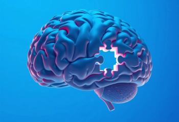
[[{"type":"media","view_mode":"media_crop","fid":"57718","attributes":{"alt":"© SHUTTERSTOCK.COM","class":"media-image media-image-right","id":"media_crop_5200317488686","media_crop_h":"0","media_crop_image_style":"-1","media_crop_instance":"7273","media_crop_rotate":"0","media_crop_scale_h":"232","media_crop_scale_w":"200","media_crop_w":"0","media_crop_x":"0","media_crop_y":"0","style":"font-size: 13.008px; float: right;","title":"© SHUTTERSTOCK.COM","typeof":"foaf:Image"}}]]
Learning neuroanatomy is difficult
What makes learning neuroanatomy difficult? First, in and of itself, neuroanatomy can be dry and boring. (Surely, I am not the only psychiatrist who finds this to be the case.) Yes, I am awed to contemplate how a mere 3 pounds of brain, the consistency of firm pudding, could possibly be the basis of who we are as human beings and also as unique individuals. It is precisely these thoughts that bring me face-to-face with one fundamental problem that many psychiatrists encounter in thinking about neuroanatomy: What does learning about brain structures have to do with what I really want to know? Indeed!
While I am very interested in the neurobiological basis of human experience, it doesn’t really matter to me whether, for example, memory consolidation or the processing of fear takes place in a brain structure called A or B. What I want from neuroanatomy are insights into behavior. Given that a person’s motivation is key to learning anything, here the psychiatrist encounters the first of many speed bumps on the road to learning neuroanatomy.
What are the other speed bumps? Undoubtedly, for anyone who has even dipped a toe into the sea of neuroanatomy, the following difficulties are likely to be familiar.
1. Neuroanatomical terminology is obscure, often deriving from Greek roots and with no modern referents to help with recall.
2. Neuroanatomical terminology is also confusing. (For example, 3 of the basal ganglia are the caudate nucleus, the putamen, and the globus pallidus. All 3, as a group, may be called the corpus striatum. Sometimes the caudate plus the putamen together are referred to as the striatum. On the other hand, the putamen may be grouped with the globus pallidus and called the lenticular nucleus. This sounds confusing because it is confusing.)
3. The underlying structural organization of the brain is not readily apparent. A variety of different approaches have been developed for defining, differentiating, and naming parts of the brain. Some of these systems are based on surface anatomical features (for example, orbitofrontal cortex, medial frontal cortex, and dorsolateral cortex), some on cellular pathology (Brodmann’s areas), others on the functional relationships of brain structures (such as the visual system, extrapyramidal system). These various approaches do not necessarily correspond directly to one another.
4. Unlike organs such as the liver, lungs, or kidneys that have repeating functional units, the brain is an extraordinarily complex organ in which each cubic millimeter is unique.
5. No part of the brain is an island, unconnected from other parts of the brain.
6. Learning to visualize the brain requires an appreciation of the relationship of various structural features in relation to one another in 3-dimensional space.
7.Increasing complexity becomes apparent as one turns up the magnification on any area of the brain. For example, in current MRI images, one voxel represents the summation of activity of over a million neurons. Another example would be the hippocampus, which is an extraordinarily complex structure, a veritable universe in and of itself, and one of only 2 regions in the brain where new neurons are generated.
8.Two neuroanatomical realities coexist: all brains have common features, and each brain is unique.
[[{"type":"media","view_mode":"media_crop","fid":"57719","attributes":{"alt":"© JOHAN SWANEPOEL/SHUTTERSTOCK.COM","class":"media-image media-image-right","id":"media_crop_5593005172380","media_crop_h":"303","media_crop_image_style":"-1","media_crop_instance":"7274","media_crop_rotate":"0","media_crop_scale_h":"159","media_crop_scale_w":"150","media_crop_w":"285","media_crop_x":"36","media_crop_y":"27","style":"font-size: 13.008px; float: right;","title":"© JOHAN SWANEPOEL / SHUTTERSTOCK.COM","typeof":"foaf:Image"}}]]9.Neuroanatomy is nonverbal, spatial information rather than verbal, narrative information. For help in learning this material, there now are numerous on-line resources and apps (such as the 3D Brain from Cold Spring Harbor Laboratory), some of which present rotating images of brain structures that facilitate appreciation of the brain in 3 dimensions.1
10. Even the best websites and textbooks of neuroanatomy are essentially schematic versions or maps, a first-level organization of vast amounts of information. A more complete picture of neuroanatomy would include the brain’s vascular structures and meninges, neuronal dendritic and axonal links, as well as the distribution of neurotransmitters and of glial cells (which are as numerous as neurons).
Benefits of learning neuroanatomy
So, why would a psychiatrist put in all the effort that is required to learn neuroanatomy?
One might be persuaded to learn neuroanatomy because the brain is the organ basis of psychiatry. Therefore, neuroanatomical literacy is part of the basic academic fund of information that contributes to one’s professional identity.
In addition, understanding localization expands one’s diagnostic acumen. While “locating the lesion” is widely recognized as a fundamental skill for neurologists, all physicians need to be familiar with the signs and symptoms of focal CNS disorders (such as strokes or brain tumors) that patients may exhibit. For psychiatrists, it is crucial to be able to recognize the behavioral manifestations of localized lesions. Word salad or Wernicke’s aphasia? Somatic symptom disorder or multiple sclerosis? Panic disorder or limbic seizures?
Knowledge of neuroanatomy also is useful and relevant if a psychiatrist wishes to understand and take advantage of the revolution in diagnostic imaging that has emerged over the past 50 years. During the vast stretch of human history, including while many of today’s senior psychiatrists were in training, the skull of a living person was a “black box” into which no one could see. Then, in the mid-1970s, computer technology applied to information from skull x-ray films made it possible to generate images of the brain itself (CT scans) for the first time. Following in succession came the development of MRI; positron emission tomography; single photon emission CT; functional MRI; imaging of amyloid deposits, tau, and other proteins; diffusion tensor imaging; and new approaches for capturing microglial activation that may provide a tool for gauging inflammation in the brain. These and other techniques serve not only to expand our diagnostic capabilities but also to open new avenues for research, including research into mental disorders.
Moreover, as the field of psychiatry evolves, biological interventions beyond ECT and psychopharmacology are becoming more widely used. Repetitive transcranial magnetic stimulation (rTMS), deep brain stimulation (DBS), and other neuromodulatory approaches to treatment are based on an understanding of neuroanatomy.
While a busy, practicing psychiatrist might opt to utilize neurology and neuropsychiatry consultants rather than expand his or her knowledge into the arena of clinical neuroscience and the relevant neuroanatomy, this knowledge is currently required of all newly graduating psychiatrists. In 2013, the Accreditation Council for Graduate Medical Education (ACGME) and the American Board of Psychiatry and Neurology (ABPN) published The Psychiatry Milestone Project2 that established competency requirements to be utilized in psychiatry residency training programs. These programs must now evaluate a trainee’s competence in 22 defined domains. One of these is the “Clinical Neuroscience” domain, defined as including “knowledge of neurology, neuropsychiatry, neurodiagnostic testing, and relevant neuroscience and their application in clinical settings.”
Deep lessons
Perhaps the best answer to the question of why a psychiatrist might want to learn neuroanatomy is that it helps with learning to think in a different way. Neuroanatomy provides an underlying matrix by which to organize our psychiatric observations and theories within a scientifically based framework.
Since the time of Paul Broca (1824-1880), an important goal of the neurological disciplines has been to map the relationship between behavior and brain. One might argue that understanding lesion localization for specific deficits such as Broca’s aphasia is not particularly relevant for a psychiatric practice given that psychiatrists tend to be most interested in more complex behavioral syndromes such as depression, autism spectrum disorder, or schizophrenia. On the other hand, the quest to localize has led to many unexpected insights into complex human behavior.
For instance, the neurological organization of language is relevant in thinking about the mental status features of many patients who might consult with a psychiatrist. Consider patients with autism spectrum disorder who are pedantic and literal; think of schizophrenic patients who are tangential or have loose associations, or who are hallucinating verbal material. Patients with learning disabilities or post-traumatic brain injuries may have trouble with a variety of different aspects of language and communication such as word retrieval or reading. An understanding of the neurological organization of language might be applied to these various disturbances, leading to insights that could be useful in formulating the patients’ difficulties.
Specifically, in the arena of language, we know that the word and grammar aspects of language are neurologically discrete from the “music” of verbal communication, called prosody. Prosody is a crucial aspect of social interaction. Prosody includes intonation, rhythm, pace, pitch, loudness, and so on; these elements communicate emotion (emotional prosody) as well as nuances of meaning (linguistic prosody). Irony, for instance, is conveyed principally through prosody. When you say something “in anger” or “with doubt” or “deceitfully,” the clues to these communications are largely prosodic.
It wasn’t until the mid-1970s and into the 1980s that emotional prosody was mapped to the right hemisphere, including regions for prosodic expression and prosodic comprehension, mirroring the left hemisphere organization long known as Broca’s area and Wernicke’s. Patients who have autistic spectrum disorder or right hemisphere learning disabilities may have trouble with understanding prosody. Patients with schizophrenia have deficits in expression of prosody. Language functions are also involved in hallucinatory experiences, whether experienced by the patient as “inner speech” or as coming from “outside.”
But, these are just the beginning outlines of the complex neurological underpinnings of language. For example, frontal lobe executive functions are crucial in organizing a narrative, sticking to the point, keeping the goal of the conversation in mind, and in monitoring whether the listener is “getting it.” Also involved in effective discourse are neurological systems that subserve empathy, that appraise and adjust to social circumstances, and that can comprehend another person’s inner experience and point of view.
Thinking about the difficulties a patient might have (with verbal communication or in other domains of behavior) in the context of a neuroanatomical scaffolding would be an important theoretical shift for psychiatry. The quest to localize has had far-reaching consequences, including providing insights into the underlying neurological parsing of complex behaviors that can lead us to think in a more scientific way about clinical phenomena. As psychiatrists, it makes sense to explore new ways to organize our clinical observations now that we are aware that DMS diagnostic categories are not homogenous and are not etiologically based.3 While “thinking in DSM” may still guide our clinical interventions, it is now possible, given what we have learned about the brain, to think about neuropsychiatric signs and symptoms in a way that is more neurologically sound. Conceptualizing observations of patients’ deficits into categories that are known to have particular neuroanatomical bases is an important goal and can contribute to an ongoing expansion of our understanding of biological contributions to mental disorders.
In order to do this, a psychiatrist does not need to master neuroanatomy or to learn neuroanatomy first. Learning this material can be an iterative process in which one starts with learning the insights neuroscience has discovered about memory, language, social cognition, executive functioning, attention, and so on. Then, hopefully spurred on by fascination, you might find that you want to look up the relevant neuroanatomy. Motivation, clinical relevance, narrative meaning-these mark the road to making neuroanatomy memorable.
In synergy with the genetic and technology revolutions, today there is arguably no more exciting area of exploration in all of medicine than neuroscience. As psychiatrists, we are experts in mind and meaning, deeply familiar with varieties of psychopathology and the breadth of human experience. Psychiatrists are standing at the doorway to the exciting world of the brain. Yet, for some practitioners, neuroanatomy is blocking them from getting through.
MORE ABOUT Barbara Schildkrout, MD
[[{"type":"media","view_mode":"media_crop","fid":"57721","attributes":{"alt":"","class":"media-image media-image-right","id":"media_crop_4440884996544","media_crop_h":"239","media_crop_image_style":"-1","media_crop_instance":"7275","media_crop_rotate":"0","media_crop_scale_h":"159","media_crop_scale_w":"150","media_crop_w":"226","media_crop_x":"46","media_crop_y":"9","style":"font-size: 13.008px; float: right;","title":" ","typeof":"foaf:Image"}}]]The physicist Steven Weinberg said, “. . . when I want to learn about something, I volunteer to teach a course on the subject.” For me, writing is a way to learn. My first project was to write about medical conditions that can masquerade as psychiatric DSM diagnoses. That led me to want to understand more about the complex process by which doctors make a diagnosis. Also, I had found that there was an active, scientific reconceptualization of many neurological diseases taking place, and I wanted to follow developments in this area.
The brain during life was no longer a black box. I began to use writing as a process by which to integrate new findings from the world of neuroscience into my psychiatric thinking. How does any particular discovery affect my long-held theoretical notions? What questions are raised by this new information? What are the implications of this new idea for my work with patients? While many readers might imagine that writing is a lonely endeavor, I find it more akin to an intense and engaging conversation about fascinating ideas.
I count myself lucky to have found a steady source of grist for my writing mill in the weekly clinical rounds of the Cognitive Neurology Unit (CNU) at Beth Israel Deaconess Medical Center in Boston. In parallel with the world of psychiatry, the field of behavioral neurology also has been grappling with how to conceptualize behavioral disturbances. Technological innovations in neuroscience have fueled research and led to new paradigms for understanding the brain and mental disease. I am grateful to have found academic settings and professional organizations such as the American Neuropsychiatric Association (ANPA) and the Group for Advancement of Psychiatry (GAP) that foster engagement with colleagues who share my curiosity and my excitement about these emerging concepts.
As a clinician, I find that intellectual stimulation helps me to bring new ideas and renewed energy to my work with patients. I turn to writing as a way to wrestle with scientific discoveries, to bring clarity to my thinking and, finally, to share my ideas with others.
Disclosures:
Dr. Schildkrout is Assistant Professor of Psychiatry, Part-time, Harvard Medical School, Beth Israel Deaconess Medical Center, Boston, MA. She is the author of 2 books, Unmasking Psychological Symptoms: How Therapists Can Learn to Recognize the Psychological Presentation of Medical Disorders and Masquerading Symptoms: Uncovering Physical Illnesses That Present as Psychological Problems.
References:
1. Cold Spring Harbor Laboratory. 3D Brain. 2016.
2. The Accreditation Council for Graduate Medical Education, The American Board of Psychiatry and Neurology. The Psychiatry Milestone Project. July 2015.
3. Schildkrout B. How to move beyond the Diagnostic and Statistical Manual of Mental Disorders/International Classification of Diseases. J Nerv Mental Dis. 2016;204:723-727.
Articles in this issue
almost 9 years ago
Psychogenic Non-Epileptic Seizures: Clinical Issues for Psychiatristsalmost 9 years ago
Update on Medical Catatonia: Highlight on Deliriumalmost 9 years ago
Depression and Anxiety Disorders in Patients With Canceralmost 9 years ago
THE QUIZ: Headachesalmost 9 years ago
Your Money or Your Life: A Reflection on the Health Care Industryalmost 9 years ago
Westworld: Hell Hath No Limitsalmost 9 years ago
Sound of SilenceNewsletter
Receive trusted psychiatric news, expert analysis, and clinical insights — subscribe today to support your practice and your patients.


