
|Articles|May 28, 2019
- Psychiatric Times Vol 36, Issue 5
- Volume 36
- Issue 5
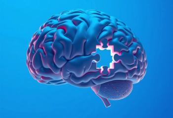
Treatment of Traumatic Brain Injury With Hyperbaric Oxygen Therapy
Author(s)Bruce I. Goderez, MD
HBOT can bring about dramatic improvement in many neurological conditions for which we have had very little to offer other than palliative care.
Advertisement
Hyperbaric oxygen therapy (HBOT) is defined as the use of oxygen at higher than atmospheric pressure for the treatment of underlying disease processes and the diseases they produce. Modern HBOT in which 100% O2 is breathed in a pressurized chamber dates back to the 1930s, when it was first used for treatment of decompression illness in divers. There are currently 13 FDA-approved uses for HBOT, including decompression illness, gas gangrene, air embolism, osteomyelitis, radiation necrosis, and the most recent addition-diabetic ulcers.
Just as practicing physicians routinely identify off-label uses for medications, over the years HBOT physicians have identified many other conditions that respond to HBOT. A number of chronic neurological conditions including traumatic brain injury (TBI) have been shown to respond particularly well. There is published literature supporting HBOT’s efficacy for TBI, including human trials and animal research, but due to the impossibility of arranging sham pressure there are no rigorous double-blind placebo-controlled trials.1 As a result, HBOT is not FDA-approved for TBI, and insurance will generally not pay for it.
HBOT can dramatically and permanently improve symptoms of chronic TBI months or even many years after the original head injury. This assertion is generally met with skepticism within the medical establishment because we have been taught for generations that any post-concussion symptoms persisting more than 6 months or so after a head injury are due to permanent brain damage that cannot be repaired. Therefore, treatment has been limited to symptom management and rehabilitative services, and any claim suggesting that fundamental healing is possible is suspect. The combination of entrenched skepticism and lack of insurance coverage has made it very difficult for patients to access treatment.
Another source of skepticism has been the large number of disparate conditions that are claimed to be helped by HBOT. A brief review of the mechanisms through which HBOT triggers healing responses, with particular reference to the modern understanding of the pathophysiology of TBI, provides a theoretical framework to explain these claims.
Physiological effects of HBOT
About 97% of the total oxygen in blood is tightly bound to hemoglobin when breathing room air (21% O2) at sea level (1 atmosphere, or 1 ATM; 3% of the oxygen is dissolved in blood serum. This amounts to about 0.3 mL of oxygen dissolved in 100 mL of serum. By the time oxygen diffuses out of the circulatory system and ultimately reaches the mitochondria, there is just a trace amount present. HBOT’s primary mechanism is to temporarily hyper-oxygenate body tissues. HBOT delivered at 1.3 ATM increases dissolved oxygen in serum by a factor of 7. HBOT delivered in hard chambers at 2.5 to 3.0 ATM increases dissolved oxygen by a factor of 15 or more. Oxygen levels in body tissues outside the circulatory system will be increased commensurately.
If a hyper-oxygenated state is maintained for long periods it will cause significant oxidative damage, but when it is “pulsed” for an hour it triggers a variety of healing processes without overwhelming the body’s anti-oxidant system. The currently known mechanisms include a powerful anti-inflammatory effect, reduction of edema, increased blood perfusion, angiogenesis, stimulation of the immune system, stimulation of endogenous antioxidant systems, mobilization of stem cells from bone marrow, axonal regrowth, and modulation of the expression of thousands of genes involved in the inflammatory response and various healing responses.2,3
Pathophysiology of TBI
At each site of impact a contusion can develop-essentially a bruise that may involve local bleeding and neuronal death. Over hours to days an area of inflammation will develop around the contusion, just as inflammation will occur around an injury anywhere in the body. Since the brain is encased in the skull swelling is strictly limited, resulting in increased pressure in the affected area. The increased pressure results in reduced blood flow, damaging a much larger area of cortex than was initially injured. Within this penumbra, neurons may be injured and unable to carry out their prime function of transmitting neuronal impulses, yet they can survive in this stunned or “idling” state indefinitely.4
This understanding of the pathophysiology of TBI explains the typical evolution of symptoms after a concussion. The patient may lose consciousness or may just feel stunned for some time. There may be an initial headache and some degree of confusion, which often improve over the next few hours. However, as the inflammatory process evolves more severe symptoms develop, usually peaking within 1 to 2 weeks. These include headache, “brain fog,” nausea, photophobia, hyperacusis, difficulty with focus and multitasking, impaired memory, difficulty with visual processing with prominent difficulty looking at screens, and profound fatigue. The symptoms eventually stabilize, then begin a slow recovery over several months.
This type of injury is referred to as “mild” TBI, since there is no gross destruction of brain matter. The absence of gross damage is reflected in the typical finding of unremarkable CT and MRI scans even in the presence of disabling symptoms. However, in many cases a brain perfusion (SPECT) scan can image macroscopic areas of reduced perfusion of the cortex.
Treatment protocol for TBI
HBOT is regulated by the FDA as a drug, and like a drug, the appropriate dose can vary with the condition being treated. Dose is determined by the pressure in the chamber and the total hours of treatment. HBOT for FDA-approved indications is most commonly delivered in hospital settings, usually in large multiplace chambers at a pressure of 2.0 ATM or higher. High pressure treatment is superior for infections and for other acute severe problems.
It took several decades to determine that, due to excessive oxidative stress, high pressure HBOT carries a significant risk of further damage in chronic diffuse neurological conditions. Treatment at lower pressures in conjunction with limits on the number of sessions has been shown to be safer and more effective for these conditions, including TBI.
The recommended protocol for TBI is currently one or more blocks of 40, 1-hour HBOT sessions delivered at 1.3 to 1.5 ATM. Treatment can be conveniently delivered in “mild” hyperbaric chambers, soft vinyl chambers limited to 1.3 ATM that are inflated by a small compressor using room air (eliminating the risk of fire). Oxygen is extracted from ambient air by a portable oxygen concentrator, removing the need for oxygen tanks. Oxygen is fed into the chamber through a tube and delivered to the patient via an ordinary hospital oxygen mask.
These chambers are affordable, simple to assemble, simple to operate, and can be used in the outpatient setting. They are considered class II medical devices similar to a continuous positive airway pressure machine, requiring a doctor’s prescription but usable at home without direct medical supervision.
CASE VIGNETTE
RP is a 55-year-old man who originally entered treatment with a 20-year history of bipolar disorder. His life had been chaotic because of lack of treatment adherence. He was stabilized on a modest dose of lithium and has been in a stable relationship and successfully self-employed for the past 10 years.
When it occurred to me to ask him about concussions, it emerged that there were several significant sports-related concussions during adolescence and at least a half-dozen serious concussions in early adulthood, possibly related to recklessness during manic episodes. His last concussion occurred 30 years before this history was obtained. He was unable to describe any specific post-concussion symptoms, possibly because he had so many concussions starting in adolescence, and could not remember what his functioning was like prior to his concussions. A brain-perfusion SPECT scan showed extensive perfusion defects consistent with TBI, which is a strong predictor of clinical benefit with HBOT. Accordingly, he was offered treatment even in the absence of a clear history of acquired symptoms.
The patient rented a mild-HBOT chamber and did the treatment at home. He completed two blocks of 40 one-hour sessions of mild HBOT (1.3 ATM, 100% O2) over the course of 4 months. A post-treatment SPECT scan was obtained about 56 months after he completed the treatment protocol.
Following is the summary section of the radiologist report for each scan. Note that a normal SPECT scan should show homogeneous perfusion, whereas areas of reduced perfusion or heterogeneous (spotty) perfusion indicate cortical areas of reduced blood flow.
Pre-treatment SPECT scan,
Aug. 29, 2016
“Findings: Decreased tracer perfusion is seen in the right temporal lobe and also there is heterogeneous perfusion in the bilateral parietal and posterior frontal lobes. The cerebellar hemispheres are symmetrically perfused in the correct clinical settings, this may reflect sequelae of traumatic brain injury.”
Post-treatment SPECT scan,
Dec. 27, 2017
“Findings: Brain SPECT images demonstrate homogeneous perfusion of the cerebral hemispheres. There are no asymmetric perfusion defects, with interval resolution of previously seen asymmetrically decreased radiotracer uptake in the right temporal lobe. Similarly, previously seen decreased perfusion in bilateral parietal and posterior frontal lobes has resolved. Impression: Normal brain perfusion SPECT with interval resolution of previously seen areas of decreased perfusion.”
After completing treatment he reported improvement in focus, improved ability to multitask, and generally more stable emotional functioning. He noted that he was using vocabulary that he had not used since he was a teenager, which was readily observable on interview. He found that he was communicating with people in a much more direct way, in contrast to his usual tendency to be tangential with difficulty getting to the point. He stopped using an appointment book for his business, finding that he could keep track of appointments with his clients for several weeks ahead by memory.
Discussion
This case is not ideal as a teaching vehicle because of the lack of clear documentation of changes in post-concussion symptoms or neuropsychological testing results. However, the normalization of a grossly abnormal SPECT scan is clear indication that HBOT can repair neurological damage even decades after an injury, bringing macroscopic areas of cortex back “on-line.” The changes in the patient’s functioning and demeanor were striking, and clinically there was no doubt about the magnitude of the response. A controlled trial in a series of similar patients including pre- and post-neuropsychological testing, rating scales, and serial SPECT scans was published in 2012.1
Cerebral palsy (CP) can be considered to be perinatal TBI and patients with CP have been shown to respond significantly to HBOT.5 Benefits brought about by HBOT in TBI and CP are generally permanent, although patients may be more vulnerable to reinjury. Clinical experience and compelling case reports suggests that Alzheimer disease and multiple sclerosis can be improved to some extent by HBOT. Benefits in patients with progressive illnesses such as multiple sclerosis will tend to deteriorate over time. A maintenance schedule of perhaps a few sessions per week can slow down and, in some cases, appears to prevent progression.6,7
Conclusion
HBOT can bring about dramatic improvement in many neurological conditions for which we have had very little to offer other than palliative care. Considering the high incidence of many of these neurological conditions, the safety of treatment, and the simplicity and relatively low cost of mild-HBOT, it is unfortunate that it is not more widely available.
Disclosures:
Dr Goderez is a psychopharmacologist and integrative medicine practitioner in private practice. He offers hyperbaric oxygen therapy for traumatic brain injury and other neuropsychiatric conditions including dementia and radiation necrosis.
References:
1. Harch PG, Andrews SR, Fogarty EF, et.al. A phase I study of low-pressure hyperbaric oxygen therapy for blast-induced post-concussion syndrome and post-traumatic stress disorder. J Neurotrauma. 2012;29:168-185.
2. Efrati S, Ben-Jacob E. How and why hyperbaric oxygen therapy can bring new hope for children suffering from cerebral palsy: an editorial perspective. Undersea Hyperbaric Med. 2014;41:71-74.
3. Harch, P. Hyperbaric oxygen in chronic traumatic brain injury: oxygen, pressure, and gene therapy. Med Gas Res. 2015;5:9.
4. Harch P, Mccullough V. The Oxygen Revolution. Hobart, NY: Hatherleigh Press; 2010.
5. Mukherjee A, Raison M, Sahni T, et.al. Intensive rehabilitation combined with HBO2 therapy in children with cerebral palsy: a controlled longitudinal study. Undersea Hyperbaric Med. 2014;41:77-85.
6. Harch PG, Fogarty EF. Hyperbaric oxygen therapy for Alzheimer’s dementia with positron emission tomography imaging: a case report. Med Gas Res. 2018:8:181-184.
7. Jain KK. Textbook of Hyperbaric Medicine. New York, NY: Springer International Publishing AG; 2017: 345-348.
Articles in this issue
over 6 years ago
Introduction: Meeting Our Personal and Professional Goalsover 6 years ago
Locum Psychiatric Practice: Unexpected, Unheralded Benefitsover 6 years ago
The Role of Social Media in Private Practiceover 6 years ago
Disability: Overview of Concepts Psychiatrists Need to Knowover 6 years ago
Introduction to Immunotherapy of Malignancies for Psychiatristsover 6 years ago
Looking for AmericaNewsletter
Receive trusted psychiatric news, expert analysis, and clinical insights — subscribe today to support your practice and your patients.
Advertisement
Related Content
Advertisement


Latest CME


Advertisement
Advertisement
Trending on Psychiatric Times
1
Depression and Long COVID Outcomes in Women: New Data Analysis
2
Artificial Intelligence, Job Loss, and the Psychiatric Significance of Work
3
Patterns of Propensity: A Review of Heinrichs’ How Psychiatrists Make Decisions
4
Investigational Psilocybin for PTSD and Ongoing Trials
5