Publication|Articles|February 12, 2026
- Vol 43, Issue 2
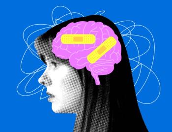
Glutamate and GABA: The Yin and Yang of the Human Brain
Author(s)John J. Miller, MD
Listen
0:00 / 0:00
Key Takeaways
- Glutamate and γ-aminobutyric acid (GABA) are key neurotransmitters, with glutamate being excitatory and GABA inhibitory, requiring precise balance for optimal brain function.
- The glutamate/GABA-glutamine cycle demonstrates an interdependence that is crucial for preventing excitotoxicity and excessive inhibition.
Explore how glutamate and GABA balance brain excitation and inhibition, shaping development, learning, and disorders like depression, schizophrenia, and autism.
Advertisement
The concept of integrating opposites or fragments into a cohesive unity is found throughout history and various cultures, with yin and yang a well-known symbolic example. The traditional symbol illustrates the interconnectedness of the masculine and feminine attributes of reality, forming a unified circle where each side contains the seed of the other as it flows into its respective partner.
Neuroscience is no exception to this concept, and it is best exemplified by the integration and interdependence of the 2 opposing neurotransmitter systems: glutamate and γ-aminobutyric acid (GABA). Glutamate is the most common excitatory neurotransmitter, whereas GABA is the most common inhibitory neurotransmitter. They both function optimally when their balance is precise, which results in harmonious neuronal function and brain health. Perturbations in either circuit can wreak havoc on brain function, and the resulting disequilibrium contributes to many major brain diseases and disorders.
When the synergistic excitatory-inhibitory balance (EIB) of glutamate and GABA is optimal, the brain is metabolically, functionally, and operationally efficient and effective at learning new information, recalling memories, processing sensory information, controlling motor movements, expressing emotions, facilitating neuroplasticity, and maintaining network stability. This EIB varies across different brain regions and during the brain’s development from birth onward. It is widely hypothesized that dysfunction in various components of the glutamate-GABA system contributes to neurodevelopmental and neurodegenerative disorders (
Glutamate
Glutamate is the most abundant amino acid in the vertebrate nervous system. Significantly, it is unable to cross the blood-brain barrier; therefore, its synthesis is required in glutamatergic neurons, which depend on glutamine transport from nearby astrocytes, the immediate precursor. Remarkably, glutamate is the direct synthetic precursor to GABA, demonstrating the interconnectedness of this vital circuitry of EIB that controls brain function.
Not surprisingly, the glutamate circuitry is quite complex and includes 3 ionotropic receptors (N-methyl-d-aspartate [NMDA], α-amino-3-hydroxy-5-methyl-4-isoxazole propionic acid [AMPA], and kainate) as well as 8 metabotropic receptors (ie, G protein–coupled receptors). The NMDA and AMPA ion channel receptors are complex, comprising heterogeneous subunits that enable a wide range of functions. Excitation of NMDA receptors by glutamate increases calcium ion flux, and activation of AMPA receptors results in sodium ion flux. Ion channels allow for rapid signaling, whereas G protein–coupled receptors provide a slower response to activation.
One important function of glutamate excitation in neurons is to unleash a cascade of events that directs synaptogenesis and neuroplasticity. If this excitation is unchecked by GABA inhibition, runaway excitation occurs, which increases the likelihood of seizures and neuronal death from excitotoxicity. As mentioned, glutamate is not metabolized in the synapse; rather, it is transported by excitatory amino acid reuptake transporters (EAAT) into adjacent astrocytes, where it is converted into glutamine. Glutamine is then transported back to the glutamate neuron by the astrocyte and converted back to glutamate. This synthetic cycle is integrated with GABA metabolism (discussed in the next section). Glutamate is primarily released from neurons by fusing intracellular glutamate-filled vesicles into the synapse, with the expelled glutamate dispersing through entropy. However, in certain physiological and/or pathophysiological states, the EAATs pump glutamate back into the synapse. For example, this occurs during cerebral ischemia.
GABA
GABA serves as the brain’s primary inhibitory neurotransmitter, and its GABAA ionotropic receptor increases the flow of chloride ions intracellularly when activated, resulting in the hyperpolarization of the neuron, which in turn inhibits neuronal firing. This is a commonly targeted receptor for the treatment of seizure disorders, anxiety, insomnia, and
There is also a GABAB receptor, which is metabotropic and the target of baclofen, an FDA-approved medication used to treat muscle spasticity. GABAB receptor activation decreases the release of both GABA and glutamate presynaptically, resulting in feedback inhibition. GABA’s inhibitory function serves many purposes, including suppressing background activity, which enhances the circuit’s signal-to-noise ratio, sharpens the signal timing, prevents overexcitation by glutamate, and maintains stability of the circuit. Significantly, GABA is synthesized directly from glutamate by the enzyme glutamic acid decarboxylase. Like glutamate, GABA is not metabolized in the synapse after release but is instead transported back into the GABA neuron or, more commonly, into adjacent astrocytes, where it is converted to glutamine.
The Glutamate/GABA-Glutamine Cycle
The glutamate/GABA-glutamine cycle nicely demonstrates the interconnectedness of the excitatory and inhibitory circuitry of the human brain (
This complex cycle of recycling the synaptic neurotransmitters glutamate and GABA is essential for optimal functioning of the circuitry to prevent excitotoxicity by glutamate and/or excessive inhibition by GABA. Significantly, this “couples” metabolism, synthetic processes, and homeostasis of the excitatory-inhibitory circuitry into an integrated and interdependent cycle in a manner reminiscent of the yin and yang archetype of ancient Chinese philosophy.3
Glutamate and GABA Cross Talk
Various types of cross talk exist between glutamate and GABA signaling, reinforcing the integration of these 2 oppositional neurotransmitters.4 A recent study identified a novel, direct interaction between glutamate and the GABAA receptor, highlighting the importance of a rapid and immediate feedback loop.5 A binding pocket for glutamate was discovered at the α+/β– subunit interface of the GABAA receptor.5 When bound, glutamate allosterically potentiates the GABAA receptor, resulting in an immediate increase in inhibitory signaling and rapid cross-talk feedback to regain balance in the circuit.5
Role in Neurodevelopment
In brain development, immature GABA neurons initially depolarize rather than hyperpolarize due to transient changes in chloride gradients, which in turn facilitate neuronal migration, growth, and synapse formation. With further brain maturation, the chloride gradients stabilize, and GABA neurons mature to their ultimate inhibitory function with the establishment of the mature EIB.
It is well established that during adolescence, the human brain undergoes progressive maturation of the prefrontal cortex, which correlates with improved cognitive development and control. Perica et al hypothesized that adolescence involves a critical period of developmental plasticity, marked by dynamic changes in the excitatory and inhibitory processes of glutamate and GABA circuitry, which typically results in normative development of the adult brain.6 Dysfunction of the EIB during this critical phase of brain development may contribute to neurodevelopmental disorders, such as
Clinical studies have utilized magnetic resonance spectroscopy (MRS) to measure concentrations of glutamate and GABA in vivo in the human brain. Steel et al demonstrated balanced excitation and inhibition in the resting human brain using MRS to quantify glutamate and GABA in brain regions.7 This is a consistent finding in the published literature and is hypothesized to be essential to maximize precise neuronal excitation timing while simultaneously preventing runaway excitation in cortical circuits via inhibition.
Disorders Likely Impacted by Glutamate/GABA Dysfunction
It is not surprising that a range of psychiatric and neurological disorders can result from wide-ranging etiologies.
• MDD—Starting in 1958 with the FDA approval of iproniazid for the treatment of MDD, all antidepressants up until 2019 worked by modulating one or several of the monoamines serotonin, dopamine, and/or norepinephrine. In the late 1990s, researchers at Yale University observed a rapid antidepressant response to a single, subanesthetic intravenous infusion of ketamine, which ignited a plethora of research that has established the effectiveness of ketamine, esketamine (FDA approved as intranasal Spravato in 2019), and dextromethorphan (FDA approved in 2022 as Auvelity, a combination of dextromethorphan/bupropion) as rapid-acting antidepressants.8-12 The putative mechanism of action of these 3 medications is antagonism of the NMDA receptor; this results in decreased excitation of GABA interneurons, which disinhibits the glutamate neuron that the GABA interneuron was inhibiting. It is hypothesized that this results in a glutamate surge, ultimately agonizing postsynaptic AMPA-glutamate receptors and triggering production of brain-derived neurotrophic factor, which orchestrates synaptogenesis and neuroplasticity (
This cascade of events is believed to be responsible for the observed increase in global brain connectivity in the prefrontal cortex of individuals with MDD within 24 hours of a single ketamine infusion and imaged with functional connectivity MRI.13,14 This putative mechanism for a rapid antidepressant effect demonstrates the complex interdependence of the glutamate and GABA circuitry in the human brain.
• Schizophrenia—Phencyclidine (PCP), also known as angel dust, is a well-established dissociative anesthetic demonstrated to function primarily as an NMDA receptor antagonist. Discontinued as a prescription drug for human use in 1965 due to severe adverse effects, PCP served as the congener for the development of ketamine, which was FDA approved in 1970. As early as 1959, articles were published describing PCP as a schizophrenomimetic drug.15 A subsequent publication, in 1962, reported that PCP administration induced positive, negative, and cognitive symptoms in “normal” participants similar to those seen in individuals with chronic schizophrenia.16 Replication of these findings led to the glutamate hypofunction hypothesis of schizophrenia, which links antagonism of NMDA receptors by drugs like PCP to disrupted cortical synchrony.
A recent review exploring evidence obtained by neuroimaging studies of individuals with prodromal, first episode, and chronic schizophrenia demonstrated regional differences in neurotransmitters, including glutamate, GABA, and dopamine. Compared with healthy controls, these individuals had higher levels of glutamate and dopamine in the basal ganglia and lower levels of glutamate, GABA, and dopamine in cortical regions, especially in the frontal cortex.17
• ASD—It has been hypothesized that EIB dysfunction may contribute to the development of ASD. Drenthen et al used MRS to quantify occipital concentrations of glutamate and GABA in 33 adolescents, 15 with high-functioning autism (HFA) and 18 healthy controls. They specifically chose to study adolescents with HFA to ensure that any observed differences from the control group were caused by ASD rather than by intellectual impairment. The researchers found increased glutamate and decreased GABA levels in the individuals with HFA vs the control group, suggesting that EIB dysfunction may contribute to atypical brain development in ASD.18
Concluding Thoughts
Glutamate and GABA, although oppositional in their neurophysiology, are intimately integrated in the human brain through their parallel distribution, interdependent metabolism via astrocytes, and codependent role in maximizing the accuracy and efficiency of the location, timing, and degree of synaptic output for the shared outcome of EIB and homeostasis.
References
1. Pavlovic ZM, ed. Glutamate and Neuropsychiatric Disorders: Current and Emerging Treatments. Springer; 2022.
2. Sears SM, Hewett SJ.
3. Andersen JV.
4. Chai A.
5. Wen Y, Dong Z, Liu J, et al.
6. Perica MI, Calabro FJ, Larsen B, et al.
7. Steel A, Mikkelsen M, Edden RAE, Robertson CE.
8. Molero P, Auba E, del Mar Unceta M, Ortuño Sánchez-Pedreño F. The modulation of glutamatergic signaling as a potential therapeutic strategy for major depression. In: Pavlovic ZM, ed. Glutamate and Neuropsychiatric Disorders: Current and Emerging Treatments. Springer; 2022:337-357.
9. Sanacora G, Zarate CA, Krystal JH, Manji HK.
10. Abdallah CG, Adams TG, Kelmendi B, Esterlis I, Sanacora G, Krystal JH.
11. Miller JJ.
12. Cabrera Abreu C, Biorac A, Baldessarini RJ, Vázquez GH.
13. Abdallah CG, Averill LA, Collins KA, et al.
14. Abdallah CG, Dutta A, Averill CL, et al.
(Thousand Oaks). 2018;2:2470547018796102.
15. Luby ED, Cohen BD, Rosenbaum G, Gottlieb JS, Kelley R.
16. Cohen BD, Rosenbaum G, Luby ED, Gottlieb JS.
17. Howes OD, Bukala BR, Beck K.
18. Drenthen GS, Barendse EM, Aldenkamp AP, et al.
Articles in this issue
6 months ago
Welcome to Our First Digital IssueAdvertisement
Related Content
Advertisement


Latest CME


Advertisement
Advertisement
Trending on Psychiatric Times
1
Preventing Alzheimer Disease After Brain Injury: Targeting TNF With XPro1595
2
Making Sense of the New Alzheimer Treatment Landscape, With Aaron Ritter, MD
3
What's New in Psychiatric Treatment? Highlights From the 2026 Southern California Psychiatry Conference
4
PET Imaging Confirms Lower Muscarinic M1 Receptor Availability in Schizophrenia
5